Angioma – Arteriovenous Malformation (AVM)
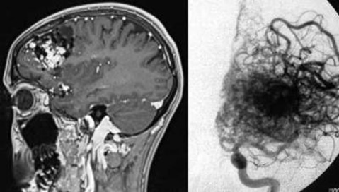
Angiomas or arteriovenous malformations (AVMs) are usually congenital vascular malformations, which consist of a short circuit of afferent vessels (arteries) and outgoing vessels (vein).
By angioma different symptoms can be triggered. In common headache, seizures and paralysis, and depending on the location of the angioma also speech or memory-efficient interference occurs. However, the main problem with angioma is a risk of brain hemorrhage occurring spontaneously. Here the risk of bleeding is specified in larger studies with AV angiomas with a bleeding probability of about 1 to 2 percent per year. For very large angiomas that have already been bleeding, the propensity for bleeding is higher.
A basic distinction between asymptomatic angiomas, ie AV Malformation who have not bled and caused no neurological deficits, as well as symptomatic angioma, for example, cause headaches, seizures or neurological deficits or have already caused a brain hemorrhage.
Following the angiographic diagnosis, arteriovenous malformation (AVM) can be due to the size, the location and the hemodynamics closer to classify (Spetzler-Martin classification). On the basis of angiography detailed planning and discussion of other methods for the treatment of angioma are possible.
In principle, three treatment methods are available:
Endovascular treatment, that is, the closure of the afferent and efferent vessels or particles with adhesive as well as with small spirals over a catheter that is introduced into the angioma
Needs: surgery, anaesthesia, hospital stay
Radiosurgery (Cyberknife), ie, targeted single irradiation of the angioma nidus, which is located in the center of the angioma bundle and thus cause irradiation-induced closure of angioma vessels and protection of the remaining cerebral blood flow in the long term.
Needs: outpatient procedure, painfree
Microsurgical treatment, that is, the complete removal of the angioma with selective occlusion of the incoming and outgoing vessels while sparing the surrounding brain vessels.
Needs: surgery, anaesthesia, hospital stay
The aim of the treatment is to close the angioma completely, because only in that case no further risk of bleeding is to be expected. The individual treatment method always has to be carefully considered, because not every angioma can be treated by endovascular or radiosurgery or surgery with the same opportunities and risks. Optionally, the combination of different treatment methods is necessary in order to achieve an angioma closure.
Since these are complex medical issues, it is important to discuss the various treatment options with a view to the individual case of disease. At the Munich Cyberknife Center the individual cases are discussed in detail with the colleagues of the stroke center of the hospital Großhadern of the Klinikum der Universität München (KUM) in the context of weekly Neurovascular conferences.


Treatment Enquiry and Appointment
Please use our contact button for a treatment inquiry and individual appointment.
Treatment Enquiry
Please use the contact button for your treatment enquiry. Your request will be answered as soon as possible.